An insulin pen is a small mechanical marvel. You dial in a dose, press the button, and a rubber plunger moves just over a millimetre forward in the cartridge. That one millimetre determines how much insulin the patient receives. But the entire mechanism is sealed inside plastic: if you want to see what happens inside, you normally have to break the pen open.
In this internal study, we used high-resolution X-ray CT to look inside a Lantus SoloStar insulin pen, before and after delivering 10 units. We measured how far the plunger moved, converted that into a volume and compared the result with the standard. The difference from the dialled dose: −3.8%, well within the permitted margin. Without disassembling a single part.
Project at a glance
| Type | Internal study, example case for medical devices |
| Object | Lantus SoloStar insulin pen (U-100), complemented by a Humalog insulin pen |
| Research question | Can X-ray CT measure the mechanical dose delivery through the displacement of the plunger? |
| Reference | ISO 11608-1:2022, dose accuracy requirements for pen injectors |
| Technique | X-ray CT scanning (Comet Yxlon FF35, helical CT) |
| Voxel size | 19.5 µm |
| Analysis | ZEISS INSPECT X-Ray |
| Result | Plunger displacement of 1.339 mm = approx. 9.62 units at a dialled dose of 10 units (−3.8%), within the ISO limit of ±1 unit |
The challenge: checking a dose you cannot see
For pen injectors, ISO 11608-1:2022 specifies how accurate the delivered dose must be. The permitted deviation depends on the dialled dose and on the dose resolution of the pen. For a pen that can be set in 1-unit steps, a maximum deviation of ±1 unit applies up to 20 units. For higher doses, it is ±5% of the dialled dose.
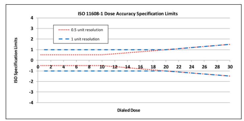
For this measurement, we dialled 10 units, or 0.10 ml of insulin (U-100 means 100 units per ml). The actual delivered dose may therefore lie between 9 and 11 units, or between 0.09 and 0.11 ml.
The problem: the parts that determine that dose sit deep inside the pen. Taking the pen apart disturbs exactly what you want to measure. X-ray CT looks straight through it. The movement of the plunger becomes visible and measurable, while the pen stays intact and can still be used.
The research question
Can X-ray CT quantify the mechanical dose delivery of an insulin pen, based on the displacement of the plunger and the geometry of the cartridge? We split that into two questions:
- Plunger displacement. Can we accurately determine the position of the rubber plunger before and after delivering 10 units?
- Volume determination. Can we convert that displacement, together with the inner diameter of the cartridge, into the displaced volume of insulin?
Our approach
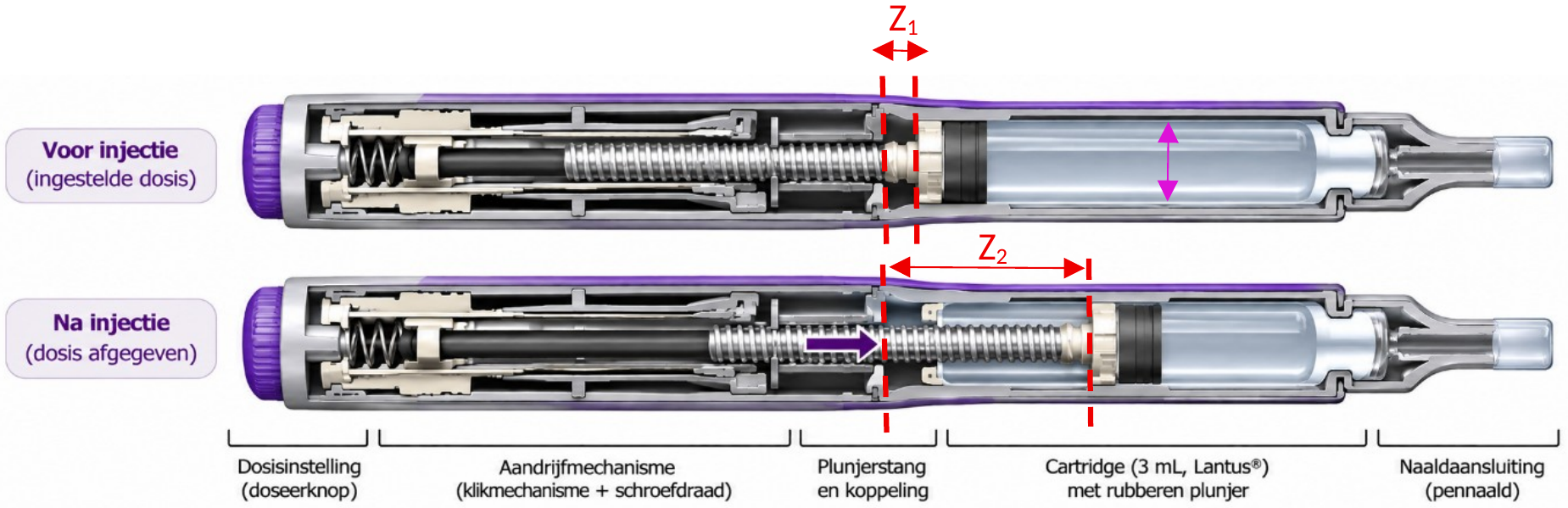
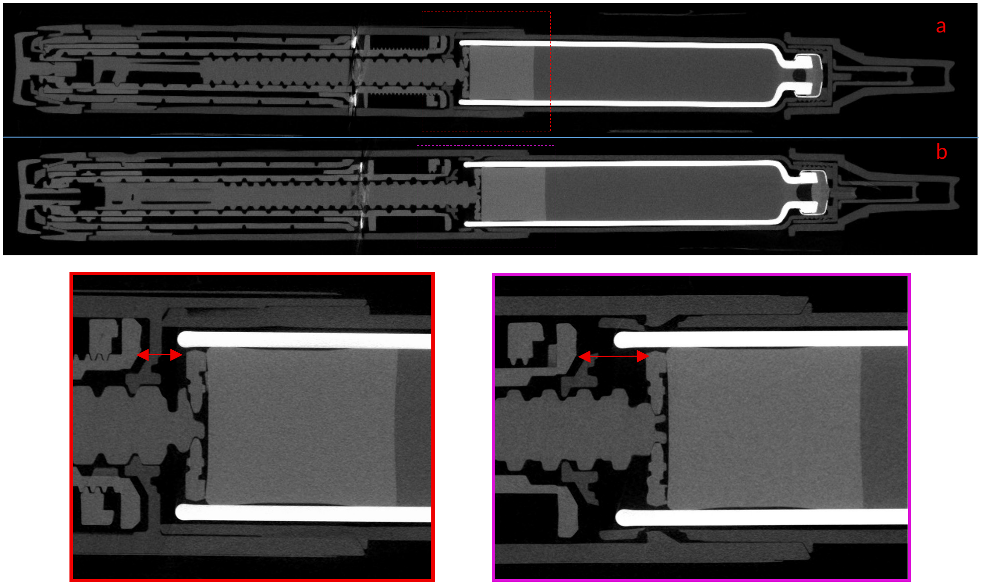
- Two scans of the same pen. We scanned the pen before the injection, then delivered 10 units and scanned the pen again. Scan, reconstruction and measurement settings were identical for both scans, otherwise you are comparing apples with oranges.
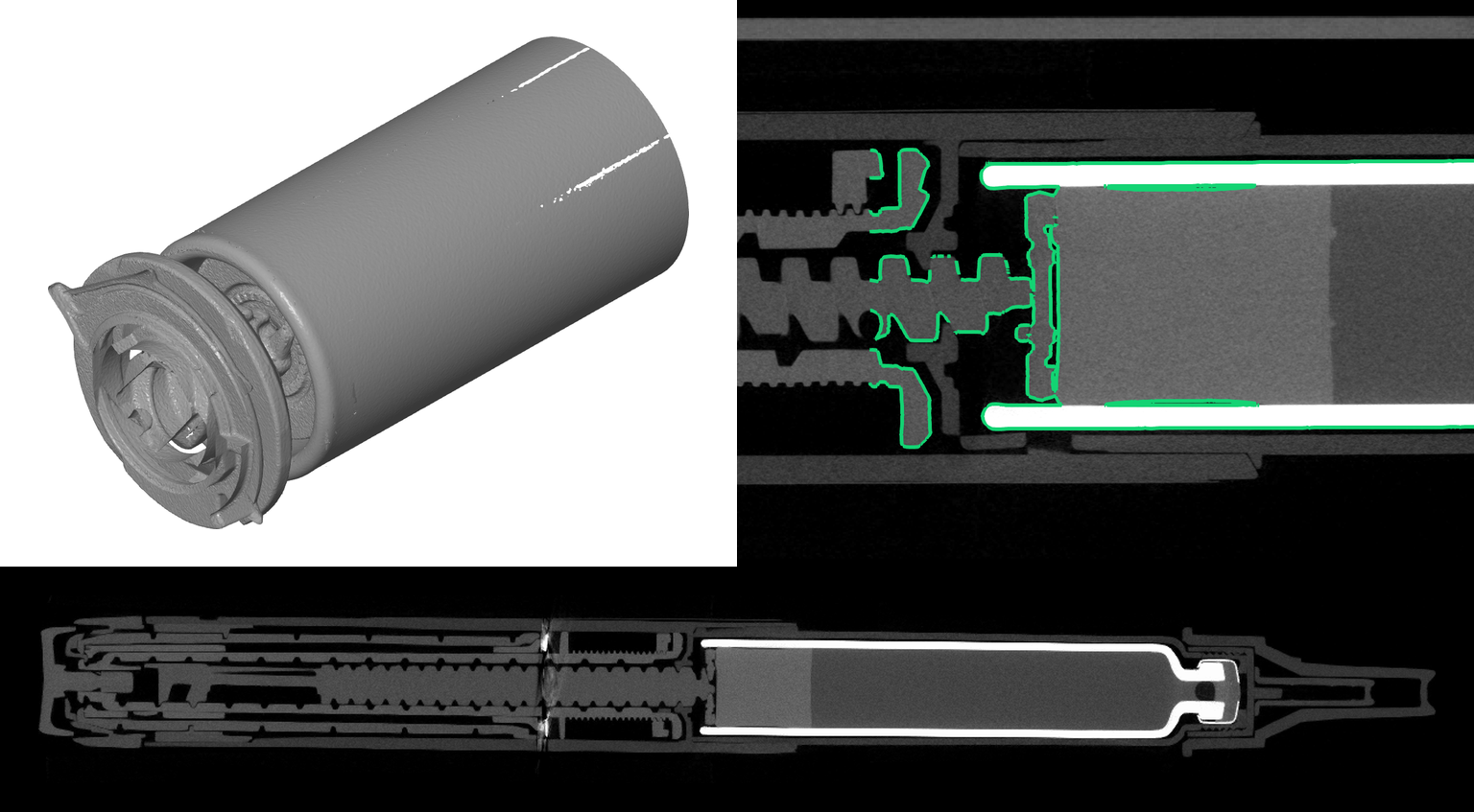
- Helical CT at high magnification. We focused the measurement on the cartridge and the plunger. With a helical acquisition on our Comet Yxlon FF35, we capture a long section of the cartridge while the pen sits close to the X-ray source for high magnification. This gave us a voxel size of 19.5 µm.
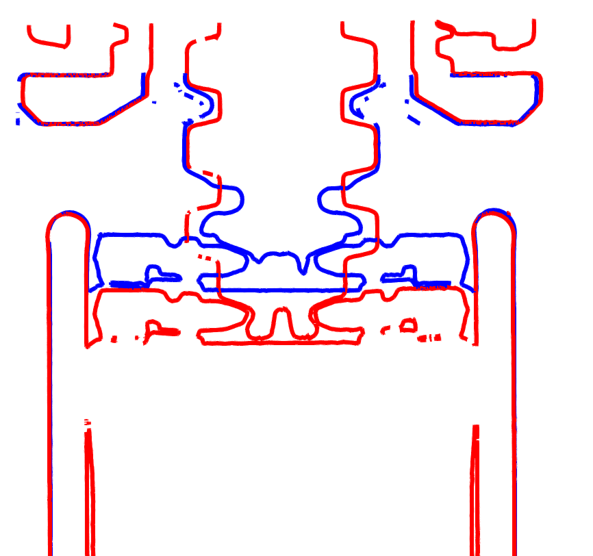
- Choosing a fixed reference. In ZEISS INSPECT X-Ray, we built a 3D model of the zone around the cartridge and plunger. That zone contains a part that does not move, the plunger holder, and a part that does, the plunger itself. The plunger holder serves as a fixed reference to align both scans.
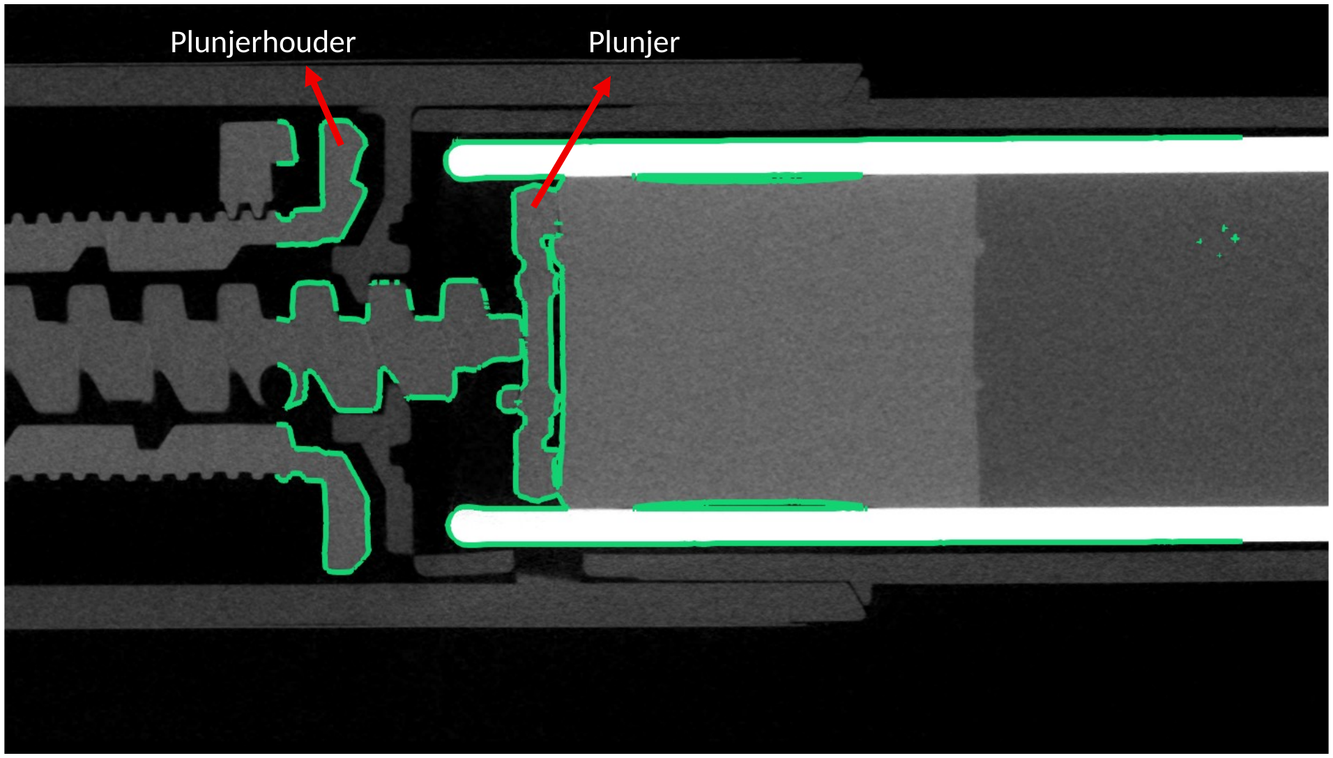
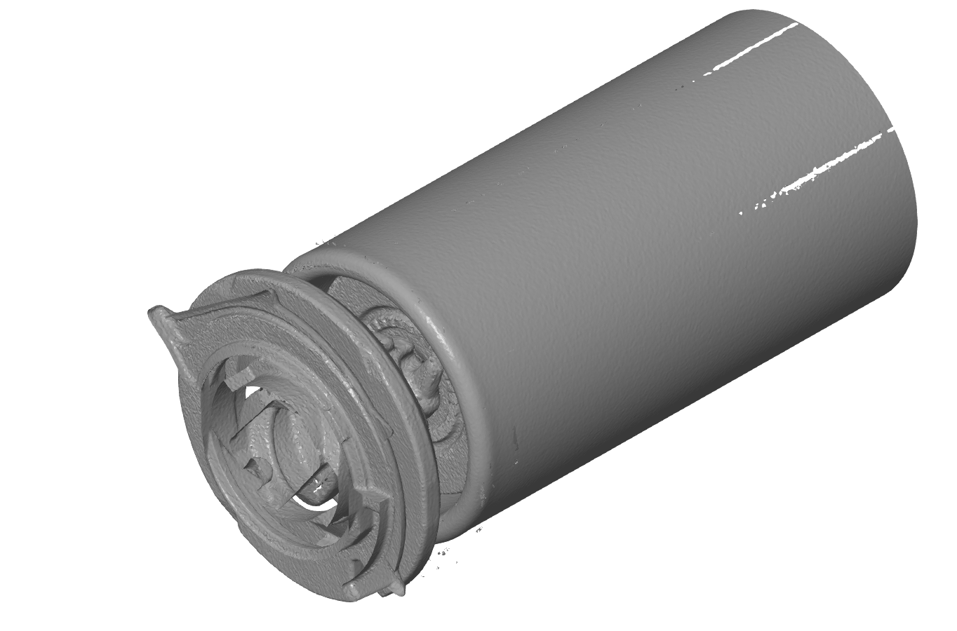
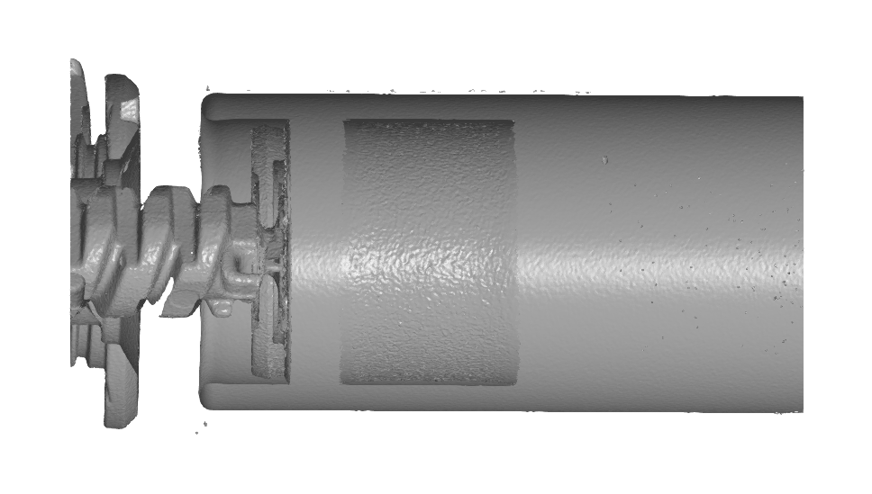
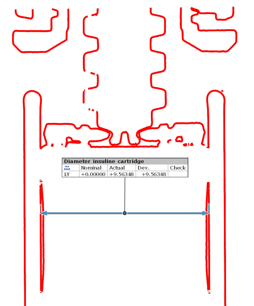
- Measuring the inner diameter. On a cross-section of the 3D model, we measured the inner diameter of the cartridge in which the plunger slides: 9.563 mm.

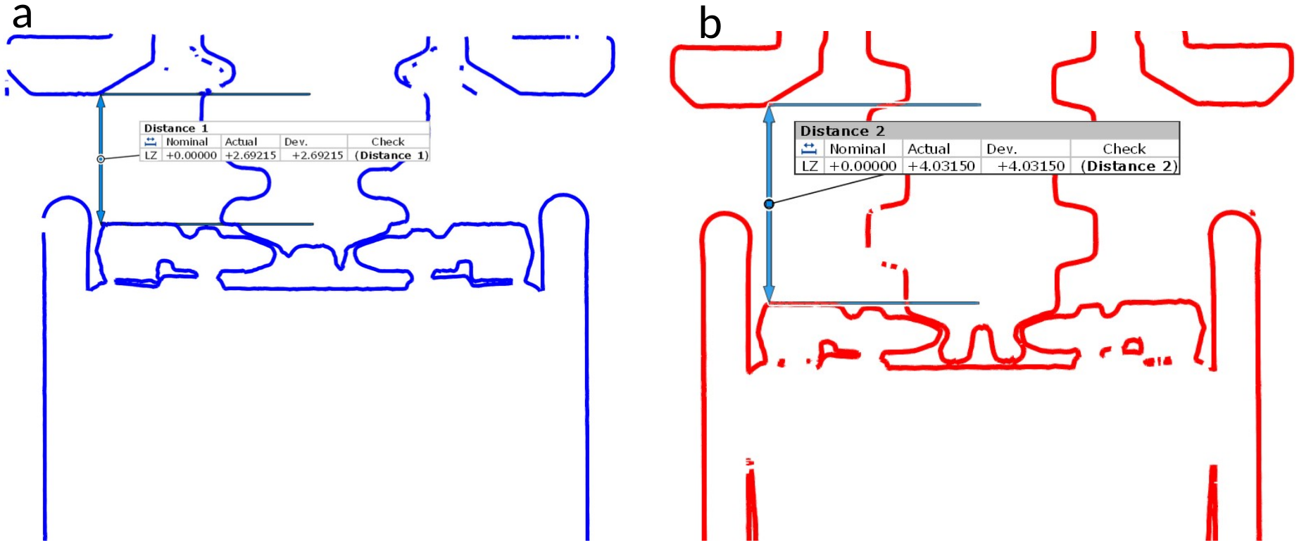
- Measuring the displacement. We measured the distance between the plunger holder and the plunger in both states. The difference is the displacement of the plunger: Δz = z₂ − z₁.
- Converting to a dose. Displacement and diameter together give the displaced volume: ΔV ≈ π · (d/2)² · Δz. We convert that to ml and then to units of insulin. In short: dialled dose → plunger displacement (mm) → volume (mm³) → calculated dose (units).
Read more about how we choose a scanning strategy for a part like this in Which CT scanning strategy is right for your part?
The result
Before the injection, the distance between the plunger holder and the plunger was 2.692 mm, afterwards 4.031 mm. The plunger therefore moved 1.339 mm. With an inner diameter of 9.563 mm, that gives a displaced volume of approximately 0.0962 ml, or 9.62 units.
| Dialled dose | 10 units (0.100 ml) |
| Distance before injection | 2.692 mm |
| Distance after injection | 4.031 mm |
| Plunger displacement Δz | 1.339 mm |
| Inner diameter of cartridge | 9.563 mm |
| Calculated volume | approx. 0.0962 ml |
| Calculated dose | approx. 9.62 units |
| Deviation | approx. −3.8% |
| Permitted under ISO 11608-1 | 9 to 11 units |
The calculated dose falls within the tolerances of ISO 11608-1:2022. More important for us: CT proves to be a suitable, non-destructive method to visualise how an insulin pen works and to measure the displacement of the plunger quantitatively.
One caveat. This study covers one pen and one dose. A volume calculated from CT is also not the same as a direct measurement of the liquid leaving the needle. It is a strong, independent check of the mechanism, not a replacement for the gravimetric test in the standard.
What else CT reveals
Besides the Lantus pen, we also scanned a Humalog insulin pen. Internally, it is built in a similar way: a large number of plastic parts around a cartridge of insulin.

At the back sits a metal spring. Metal causes typical artefacts in a CT scan, so the surrounding plastic parts appear less sharp locally. Even there, the scan still contains a lot of useful information.
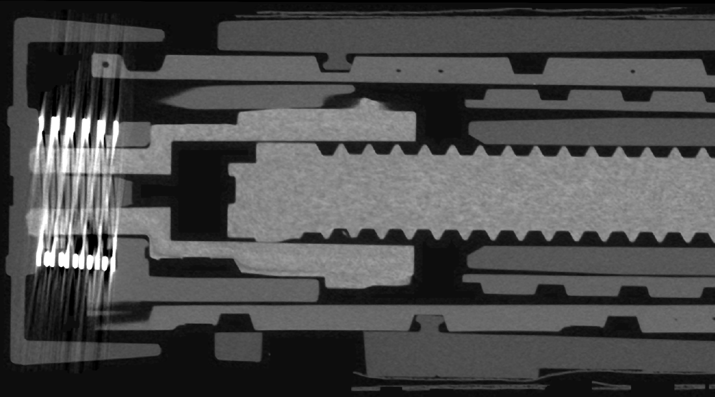
In the middle section, you can see how the plastic parts interlock and work together.
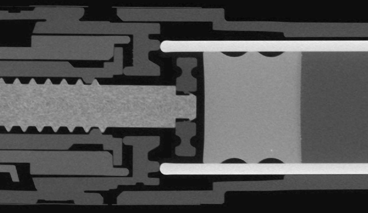
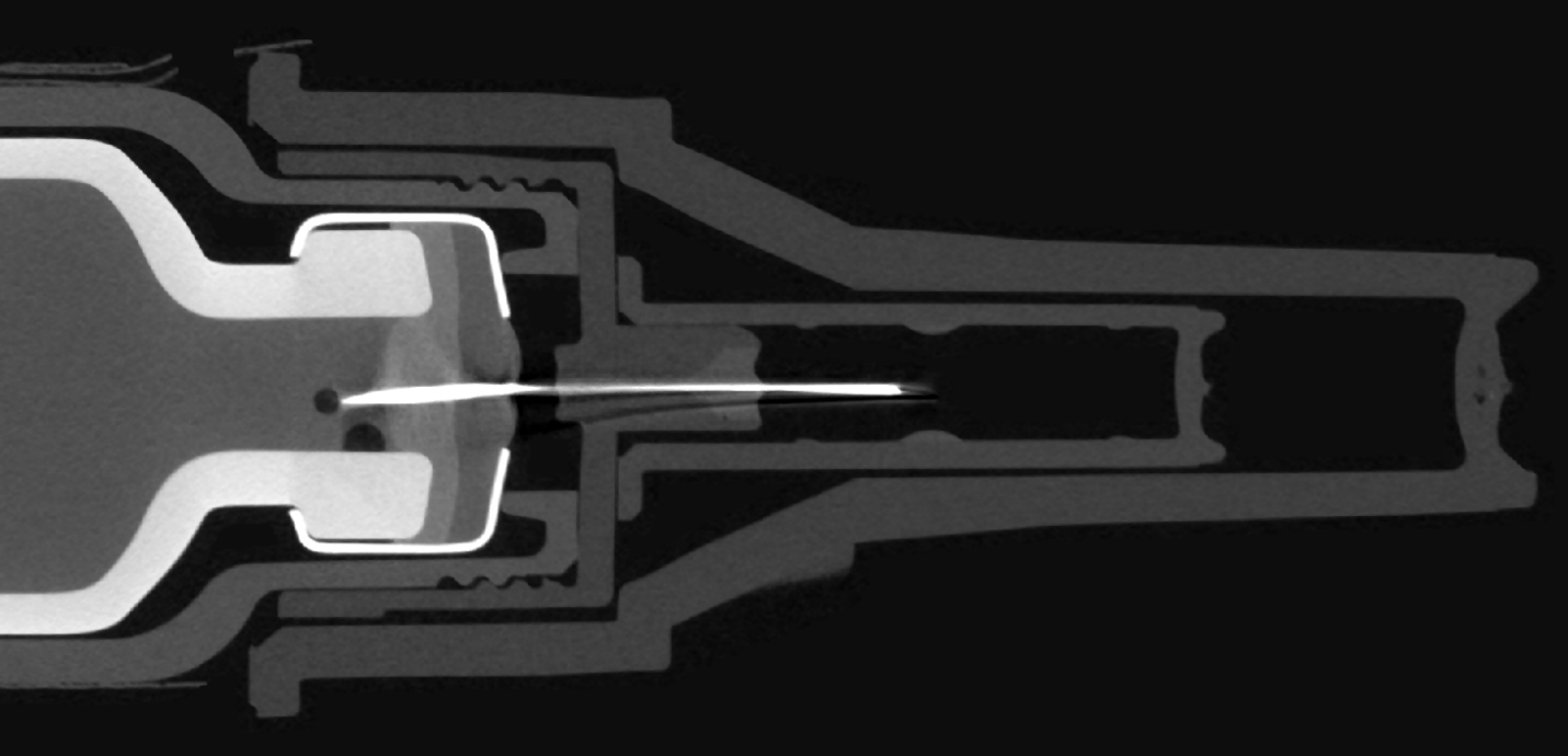
At the front, the details of the cartridge and needle come into view, and even a few small air bubbles in the insulin.
Why this matters for medical devices
An insulin pen is just one example of a closed dosing system. The same approach works for other medical devices where small mechanical movements make a big difference. We use industrial CT there for, among other things:
- non-destructive inspection of complete assemblies
- dimensional metrology of internal parts
- functional analysis, such as the position of parts before and after use
- volume determination in cartridges, reservoirs and channels
- quality inspection and comparing production batches
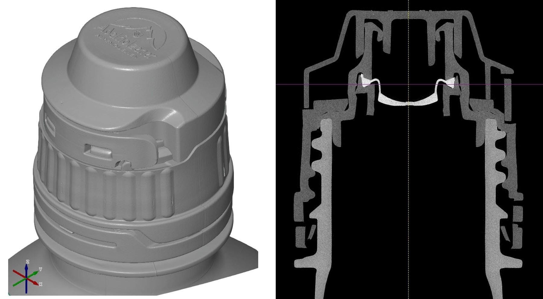
In an earlier case, we used the same approach to reveal a leaking valve system in a bottle, again without disassembling anything.
Working on a medical device?
Do you develop or manufacture injection systems, inhalers, dosing pumps or other medical devices, and want to know what happens inside? We are happy to help you find the right scan and analysis. Contact us at info@tetravision.be or +32 16 91 04 20. We always respond within 24 hours.
Want to know more about our CT capabilities? Visit our X-ray CT scanning page.