Industrial micro-CT scanning is a non-destructive imaging technique that uses X-rays to create detailed 3D representations of the internal and external structures of objects at micrometer-level resolution.
General
2D Planar X-ray images are captured and reconstructed into 2D cross-sectional slices. These slices are further processed into a volumetric data-set (3D model). The interior volume of the object is sampled in units called voxels, and a voxel can be seen as a 3D pixel.
The technology is widely used in industries like manufacturing, materials science and aerospace for quality control, defect detection and structural analysis.
Different materials have a different density, which is represented by different grey tones. When zooming in on a part you can focus on the desired area of interest, and highlight and analyse any particular feature.
Transmission of X-rays
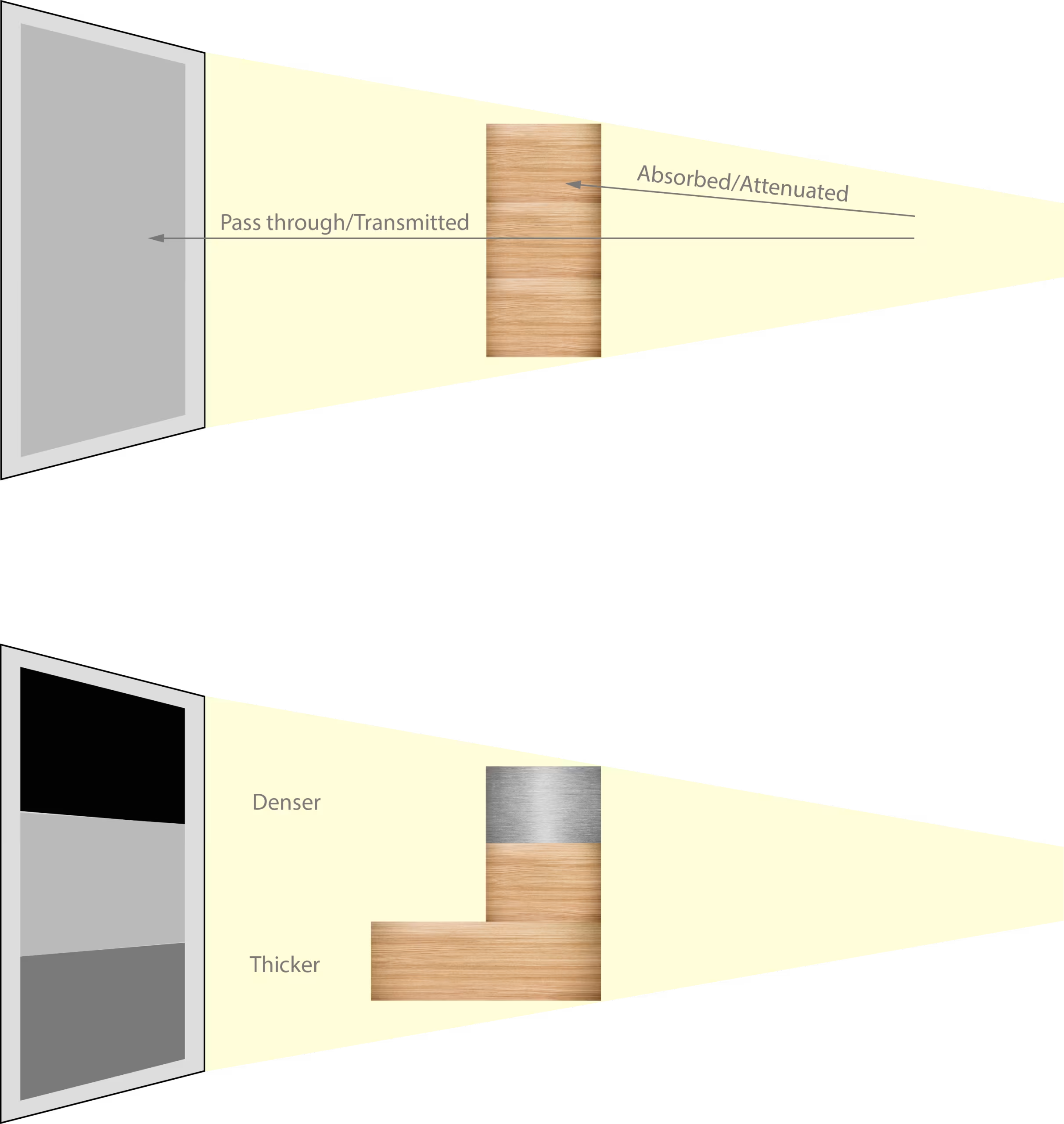
As X-rays pass through an object, some are absorbed or attenuated, while others pass through or are transmitted.
What gets through can be thought of as a shadow. The material properties of the object affect attenuation.
- The denser an object, the more X-rays will be absorbed.
- Thicker objects also absorb more X-rays.
By making different images at many angles, the scanner will be able to tell the difference between a denser material or just a thicker material.
Afterwards the collected image-data is reconstructed into a stack of tomographic images (slices). During this reconstruction the values are inverted so that air will become black and the denser objects are lighter.
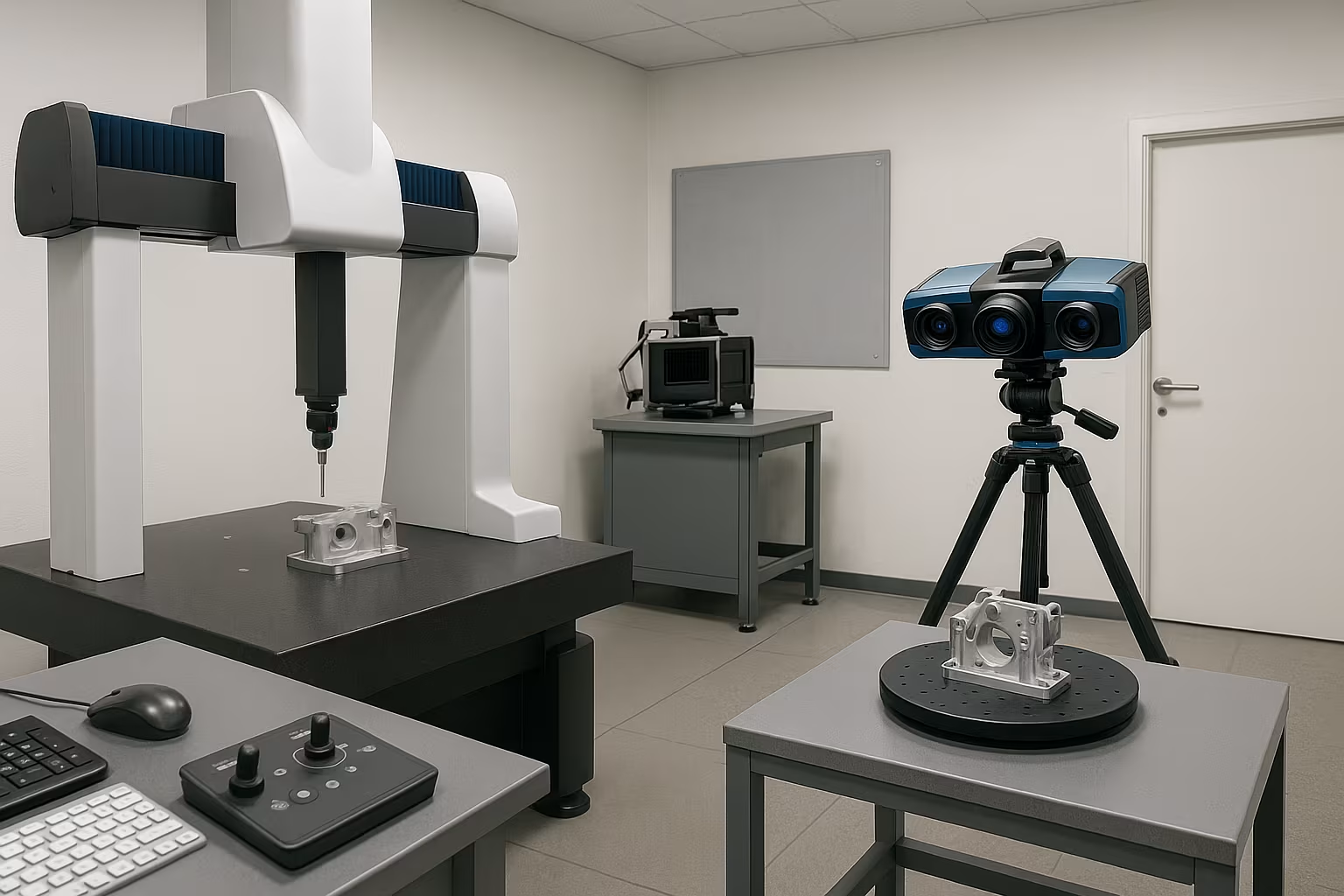
Machine principle
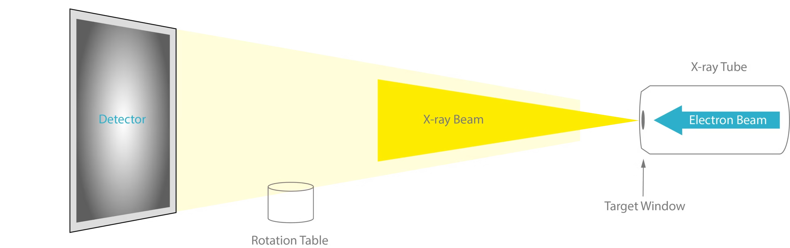
The three main parts of the CT Scanner are:
- The detector
- The rotation table
- The X-ray tube
Different types of tubes are possible, but let’s focus on a transmission tube. Here an electron beam will pass through a target window generating X-rays. Thanks to the possible movement of the part it can be positioned closer to the target or light sources, which allows a greater magnification.nen worden gepositioneerd, waardoor een grotere vergroting mogelijk is.
The power of the beam can be split into 2 components:
- Voltage
- Current
The voltage controls the energy of the photons being emitted. The more energy a photon has, the more easily it can pass through dense objects. The beam is polychromatic, which means it has a number of wavelengths and so not all photons have the same energy level. The voltage controls the maximum energy level, but less powerful photons will also be part of the beam. These photons will behave differently.
Higher energy ones will pass through the dense objects, while lower energy ones will be stopped at the surface. Since the detector panel cannot tell the difference it can create beam hardening artifacts.
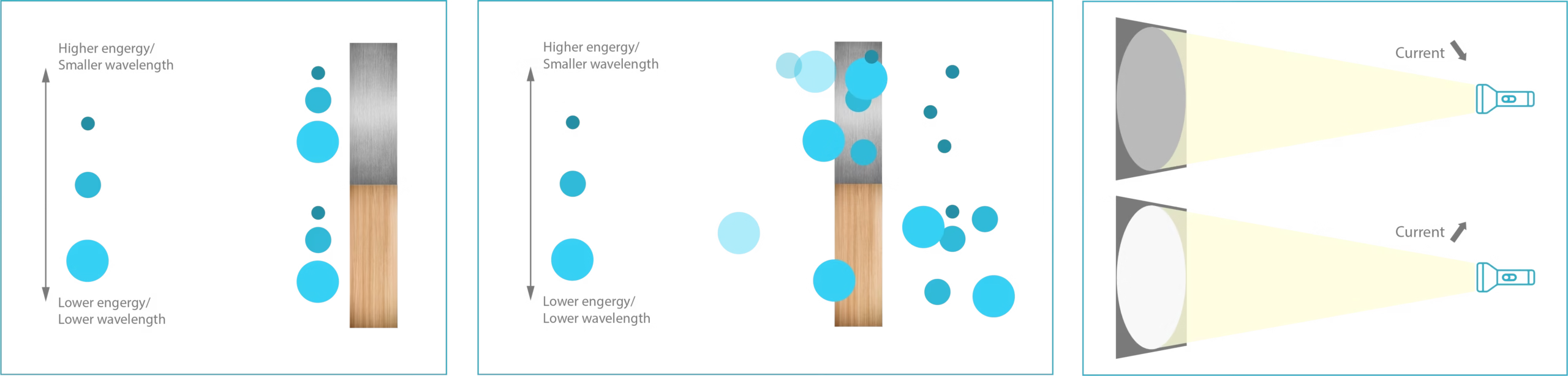
The current controls the amount of photons that are emitted. You can look at it as the brightness of the beam. Increasing the current increases the signal of the beam.
The rotation table
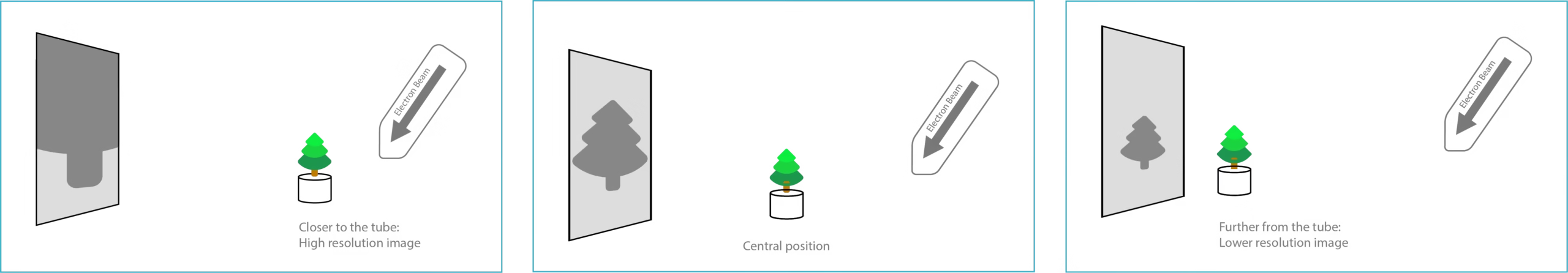
The rotation table is the base of the holder for your parts, which can be moved horizontally, vertically and in the direction of the cone beam. The distance between the part relative to the detector and X-ray tube determines the magnification and spatial resolution of the scan.
- If the rotation table is closer to the X-ray tube, it will cast a larger, higher resolution image on the detector panel.
- If the rotation table is further away from the X-ray tube, it will cast a smaller, lower resolution image on the detector panel.
Resolution is a way to describe how much details there are in a given area. This is referred to as voxel size, which is the actual amount of space represented by each voxel. For most of the industrial CT-scanners the voxels are isometric, which means they are cubic with equal X, Y and Z dimensions. If you would like to find out more on this topic, you can always check our blogpost on Resolution vs. Accuracy.
Example:
If the detector is 2000 x 2000 pixels, and the object you’re scanning is 200mm tall, each pixel will be equivalent to 100 microns. These pixels will get reconstructed into voxels of the same size.
=> 200mm/2000 pixels = 0.1mm = 100µm
If you now zoom in for a higher resolution scan, with a field of view (height of the object) of 100 mm, the voxel size decreases to 50 microns.
=> 100mm/2000 pixels = 0.05mm = 50µm
Higher resolution = Lower voxel size
The detector
The detector panel records the projected images. It can alter the acquisition time to capture a single image. A longer timing allows for more photons to be captured, and increases the signal without increasing the power of the beam. Same beam, better signal.
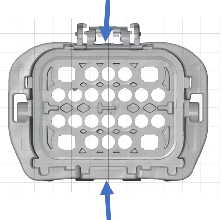
On the image on the right you can see an example of an X-ray projection on the detector plate.
Quality – Speed – Resolution
Defining the desired scan parameters requires balancing a series of trade-offs between quality, scan time and resolution. The most important aspects that have an impact on the quality are:
- Contrast: This can be improved by a high current and a good signal.
Voltage, current and timing all increase signal.
- Noise: This can be improved by capturing multiple projections and averaging them together. The more averages, the less noise.
- Artifacts: These can be reduced by using filters. Filters will reduce beam hardening, but increase the amount of noise in a scan.
Several things that can improve the quality will also increase the scan time.
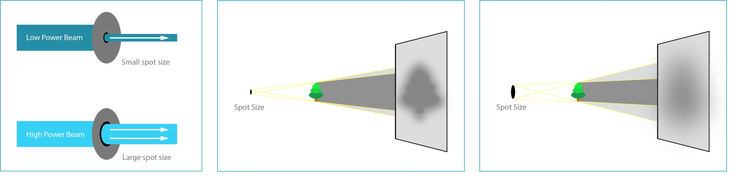
Resolution can also limit the power of the beam. As power increases, the spot size of the beam source also increases. It would be to dangerous to force a strong beam through a narrow spot size. Therefore the spot size automatically increases as power increases.
- When the spot size is smaller than the object, there is little to no blurring on the shadow that is casted on the detector.
- When the spot size increases, the image will become blurry. Moving the object away from the source, so decreasing the resolution, will help with the amount of blurring.
So to summarize: The higher the resolution is, the more limited the power of the beam is in order to get a sharp image.
If you are looking for a high resolution scan, the maximum power you can use without getting a blurry image is reduced. Reducing the power also reduces the signal and the only way to increase the signal, without increasing power, is to use a longer capture time. This will increase the length of the scan. Meaning that, indirectly the resolution can affect the scan time.