Invisible history
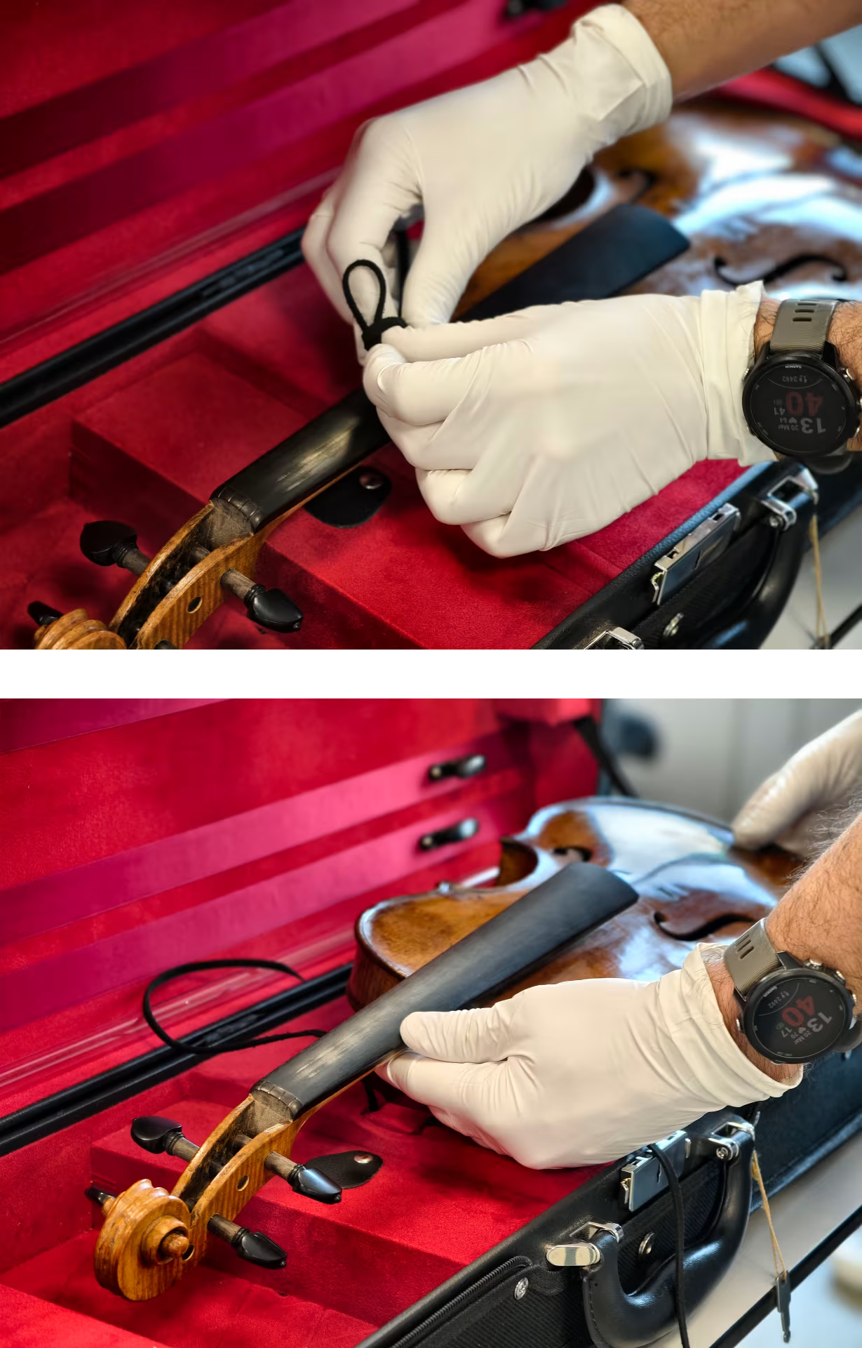
A whole new world opened up to us when we were contacted by Matthijs Strick, a renowned violinmaker at MaisonBernard. Craftsmen that go the extra mile!
Only the best is good enough, and what better way to prove the quality of their meticulous (restoration) work then showing this with actual X-ray CT-images.
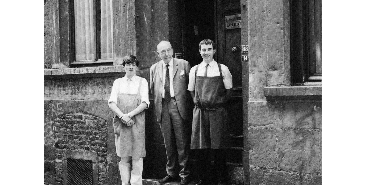
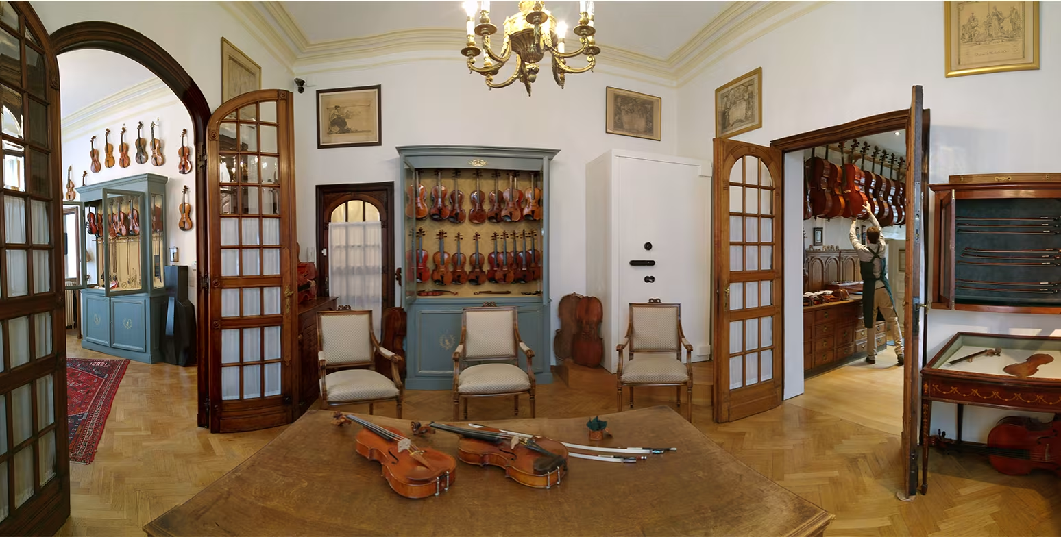
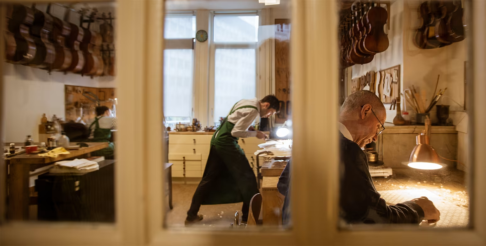
Introduction to Maison Bernard
Since 1868, Maison Bernard has been attending upon a wide clientele of professional musicians and amateurs of fine bowed instruments. Violin maker Jan Strick and Bow maker Pierre Guillaume, both appointed experts to the Prosecutor’s Office in Brussels, have been running together the oldest lutherie workshop in Europe.
Settled in Brussels since 1986, they offer a constantly renewed, extensive and unique collection of violins, violas and cellos and their bows. Jan Strick and Pierre Guillaume have earned the trust of important foundations worldwide, of collectors and some of the greatest soloists who enjoy visiting their workshop.
More info can be found on their website: https://www.maison-bernard.com/en/
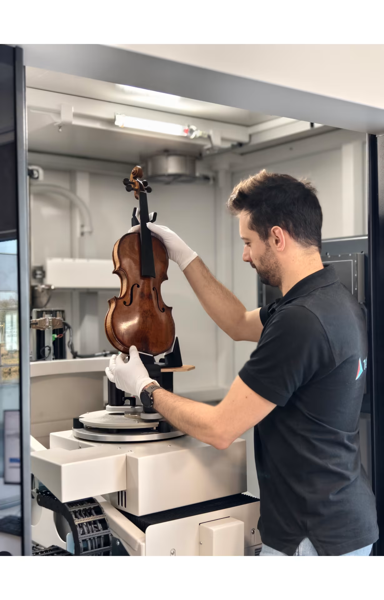
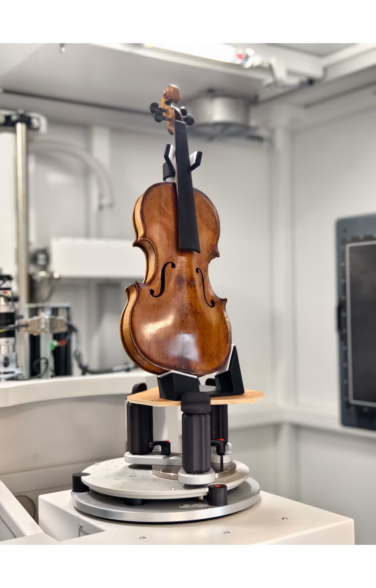
Industrial X-ray CT for violin makers
In the last decades Industrial CT-scanning has become increasingly employed in the documentation of items representing our cultural heritage. By means of CT-scanning the condition and authenticity of collectable violins can be revealed. It is a very powerful tool in both preserving and authenticating historic bowed stringed instruments. It enables detailed internal analysis beyond what visual inspection can provide.
CT-scanning also gives you the opportunity to create Finite Element Models. This way you can easily simulate and study the effect of (even minor) structural changes in the design, which can have an enormous impact on the sound.
The value of an antique violin depends on a variety of factors including:
- the condition,
- the authenticity,
- the amount of restoration applied to it,
- its lineage (was it owned be a famous musician previously).
Since X-ray CT-scanning is non-destructive and allows imaging of interior details without causing damage, it is ideally suited to deeper investigation of such objects. Let’s take a closer look and see which mysteries can be revealed!
CT Results
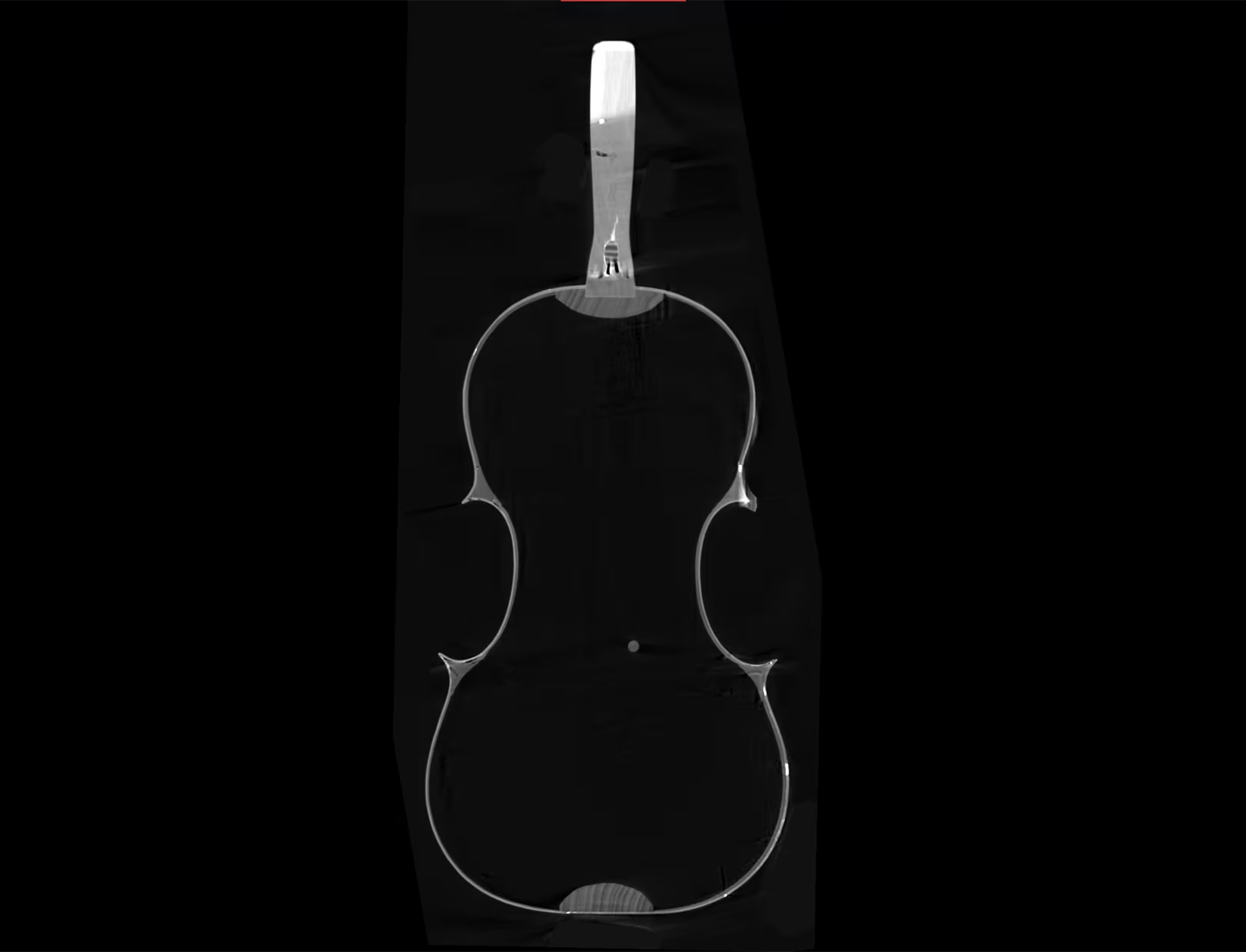
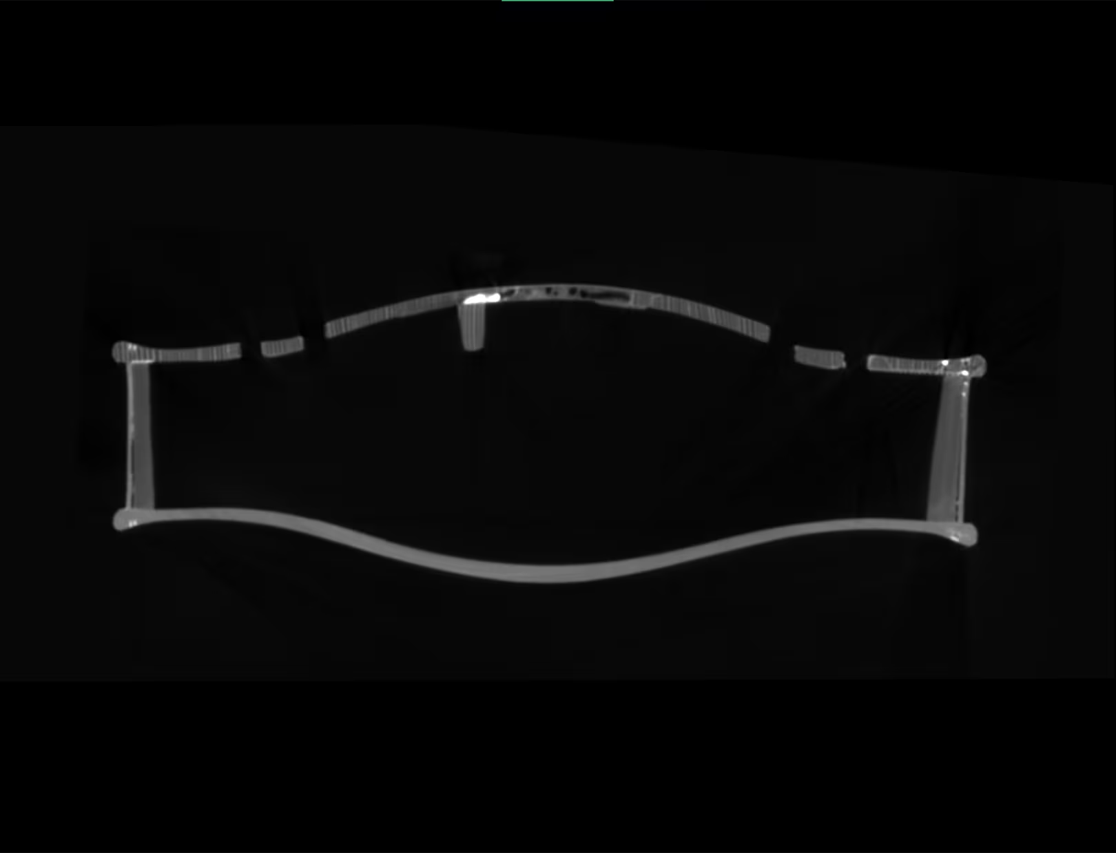
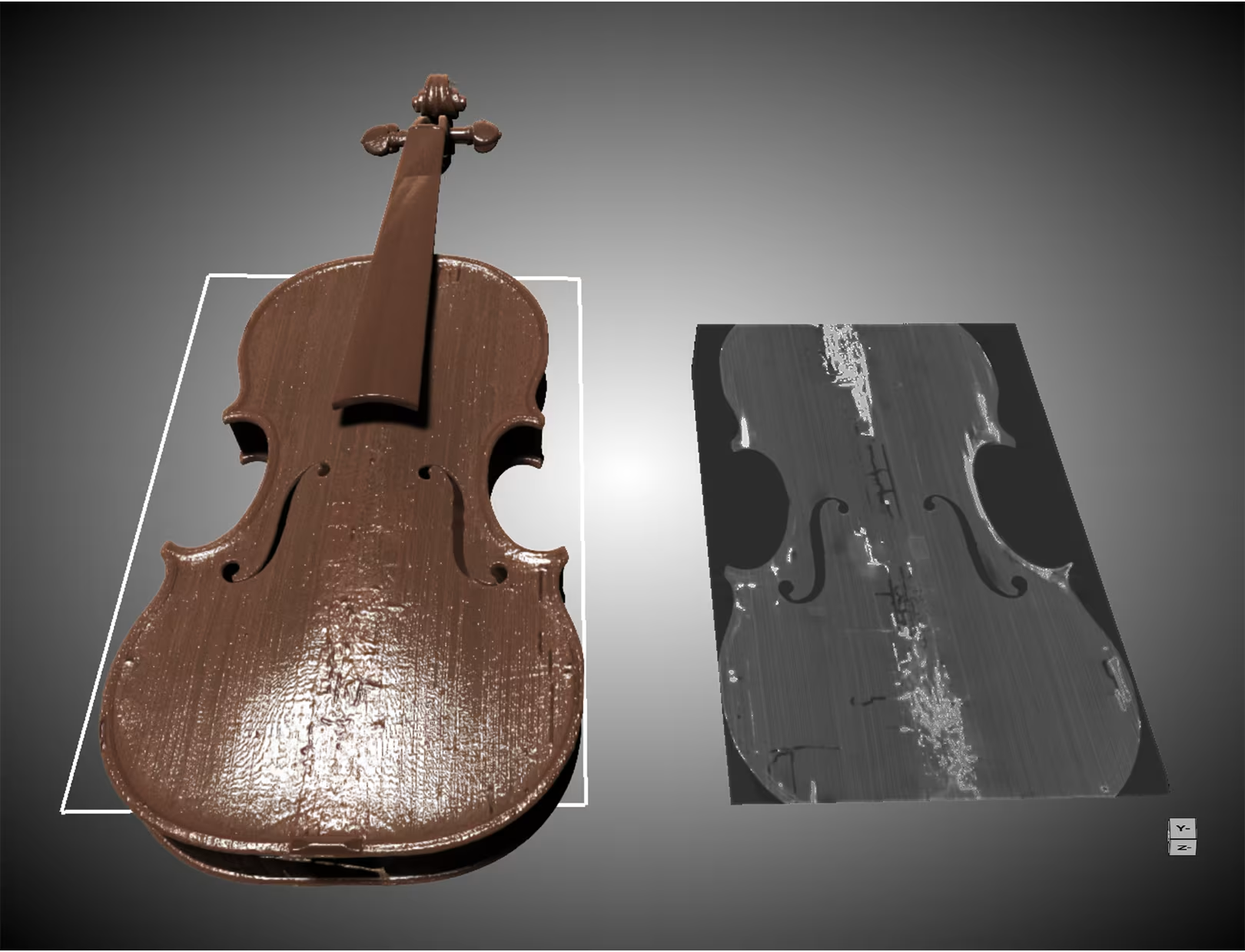
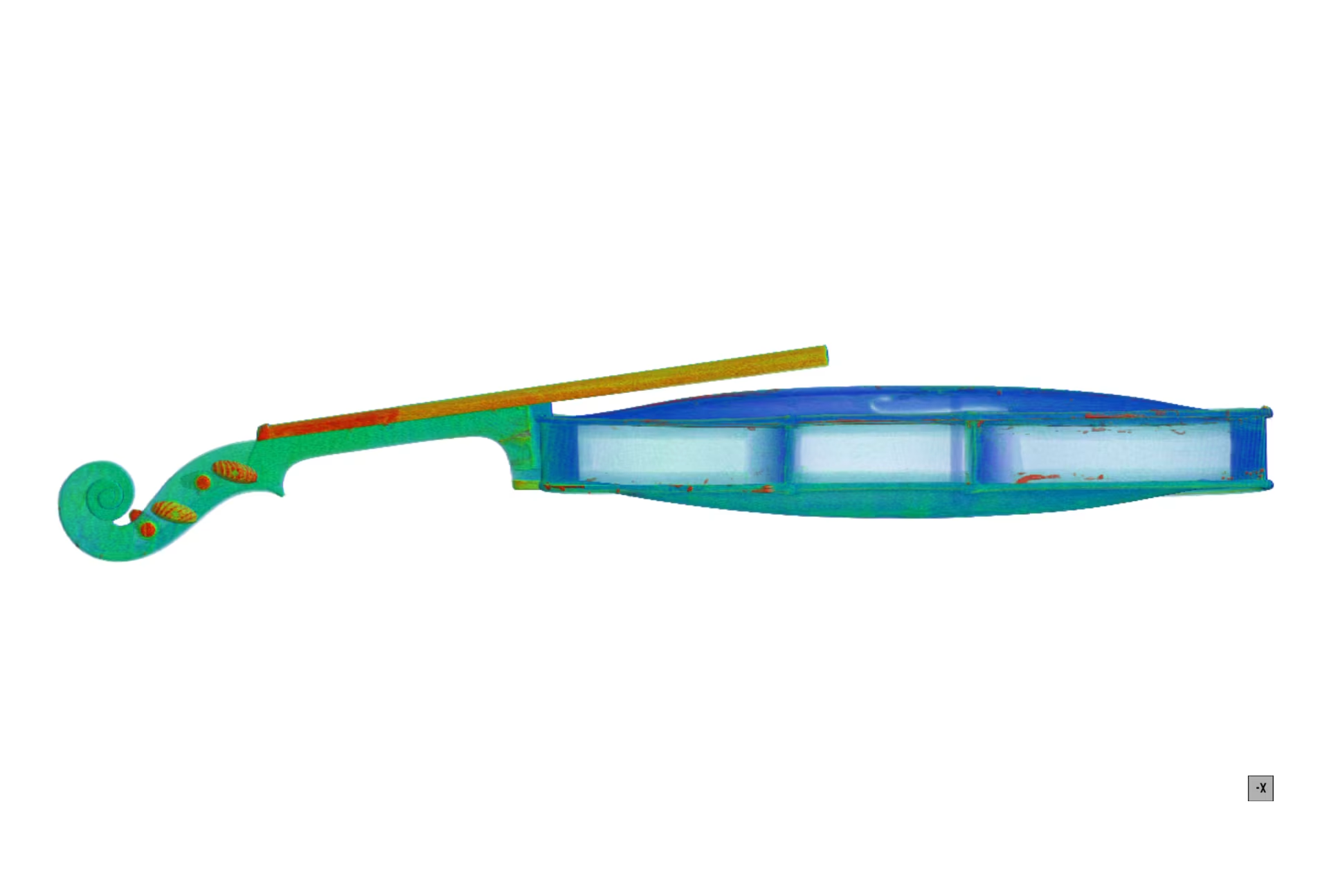
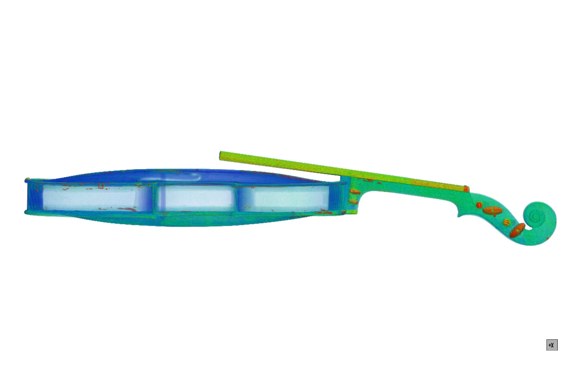
We made the CT scan using our high resolution transmission tube system. The cross sectional view in Figure 1 is parallel to the length of the violin, taking the orthogonal view to this gives a view of the curvature of the violin front face, and shows some internal details in the wood.
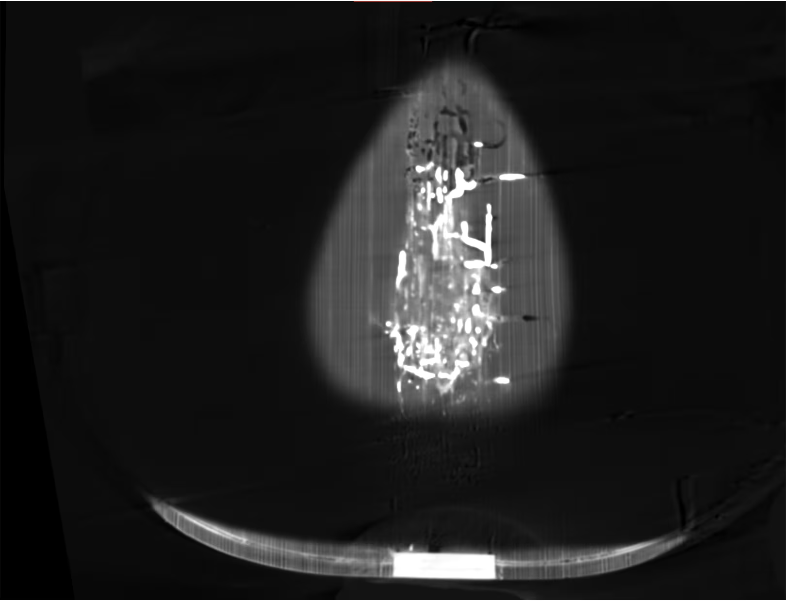
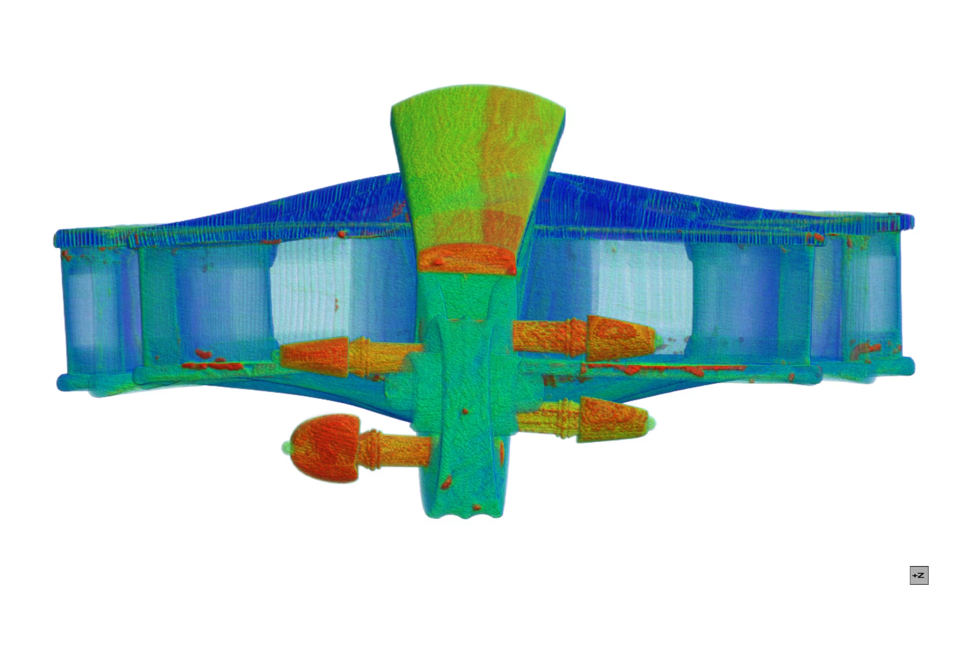
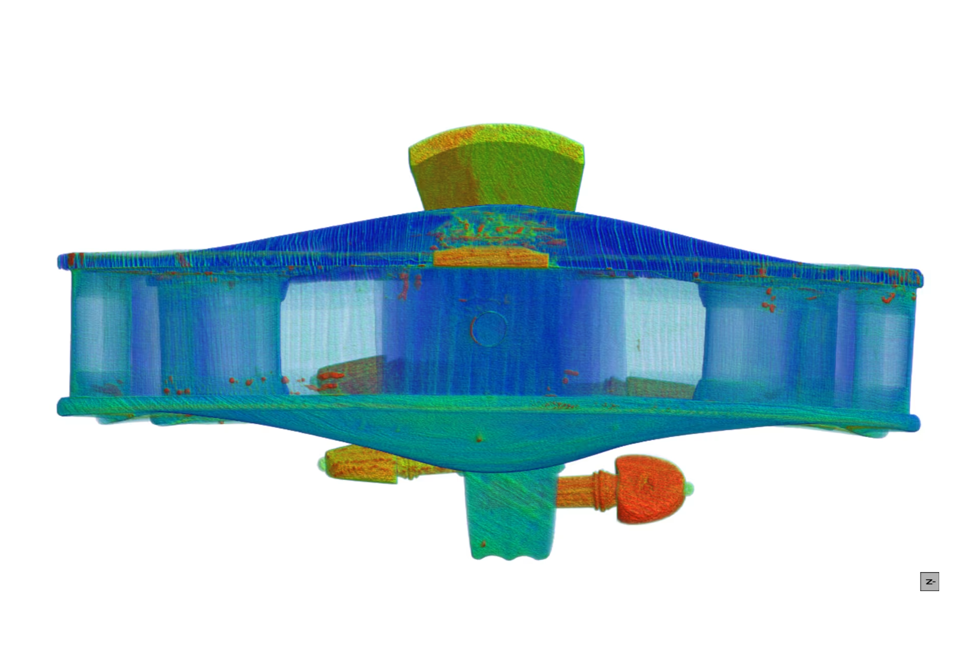
Figure 2 is showing the wood grain, as well as some interesting other details near the top of this image: what seems to be hollow channels (black) and filled channels (bright), in addition to other areas of brighter material near the edges/corners. These channels seem to be due to wood boring insects, and the filled channels indicate some effort of restoration (filler material/glue being brighter).
Dragonfly inspection
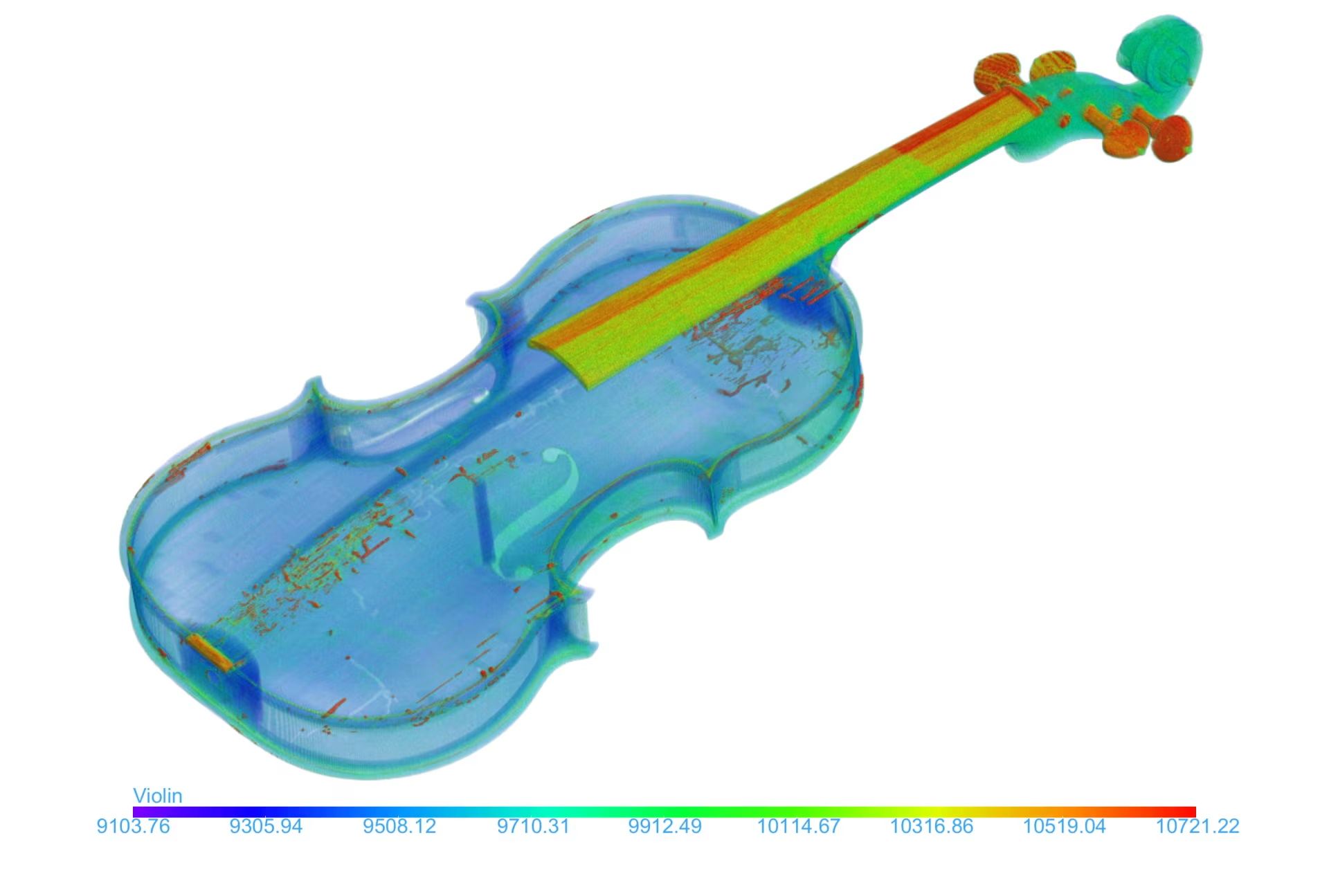
Further inspection using oblique views in Dragonfly, can help to align the cross-sectional slice with the bright and dark channels. This is shown in Figure 3.
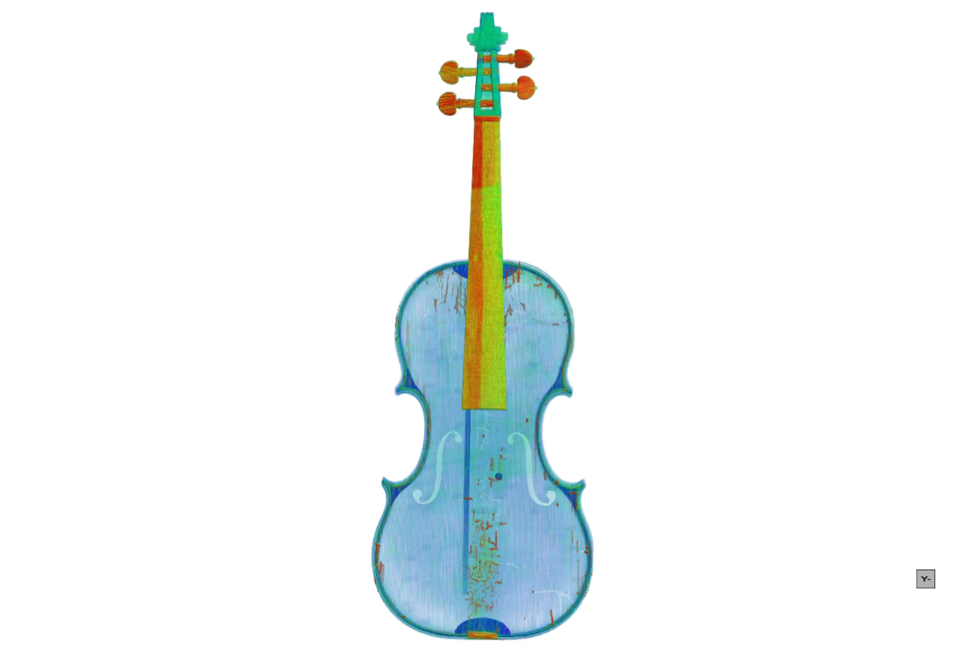
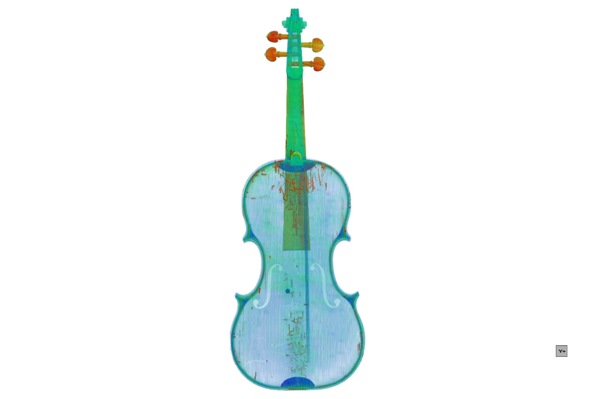
Another useful tool is the de-warping capability to manually select dewarping points in a grid, and thereby create a de-warped plane, as shown in Figure 4. This allows to see the whole front face of the violin in one cross sectional view despite it not being a planar surface.
The interesting discovery of these channels can be further quantified and visualized. Segmentation was achieved with Dragonfly’s deep learning tools, and the resulting segmentations are shown in Figure 5 in 3D views. Open/empty channels and void spaces vs. denser filled channels and other denser inclusions.
The contour dense sections are likely glue due to restoration works, but could aslo be due to the original manufacturing.
The resulting segmentations can be quantified relative to the total volume of the violin wood: for the open channels and pore spaces, the volume fraction is 0.5 %. The filled channels and glue / dense inclusions contribute 2.62 %. These type of results could be used for valuation purposes for rare antiques.
Key findings
CT SCANNING BENEFITS:
- Non-destructive detection of internal damage (e.g., wormholes, air gaps, deformities) and repairs (e.g., glue lines, filler materials, wooden cleats and patches).
- Accurate measurement of wood thickness.
- Inclusions and dense parts could be segmented to highlight previous restorations, including filled insect channels
- The inclusion % and pore space % could be quantified and potentially used as part of a quality inspection criteria for antique violins.
- Dewarping of the front face of the violin could be used to better image the channels and restored/filled channels
- Visual documentation of internal wood grain patterns unique to each instrument, aiding in authentication. CT scans provide a “fingerprint” of the instrument’s internal grain pattern, useful for identifying instruments in cases of loss, theft, or forgery.
POSSIBLE DAMAGES & REPAIR FEATURES THAT CAN BE IDENTIFIED:
- Worm infestation.
- Air gaps between structural components.
- Plastic deformities due to age and string tension.
- Cracks and extensive filler material in structural repairs.
- Animal glue lines.
- Embedded wooden cleats and patches.
- Discontinuity in wood grain indicating added material.
- …
APPLICATIONS:
- Useful to luthiers, instrument buyers, insurers, and collectors.
- Enables preservation of historical instruments and accurate reproduction by modern craftsmen.